NEURO

"Oh my neck!" Management of a cervical dislocation over 100 years ago.

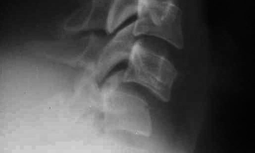
Modern radiograph of a patient with a C6-C7 cervical dislocation.

Patients sustaining cervical spine dislocations or fractures are usually immobilized at the scene with a cervical collar and a backboard and transported to a medical facility where they undergo cervical X-rays, a cervical CT scan, and possibly a cervical MRI. Treatment frequently consists of cervical reduction using traction followed by surgical fusion. Case reports on the treatment of cervical spine injuries over 100 years ago are rare. Thus the importance of the detailed case managed by Balfour Fergusson, MD, and published in the Lancet on July 8, 1899:

"At midnight on June 17th I was called to see a laborer. It appeared that he was working away from home and was in the habit of returning every Saturday night. On the evening in question, he was returning home in charge of a horse and cart and, probably overcome by a week's hard work in the hayfield, he fell asleep while sitting on the edge of the cart." Two men found the laborer lying on the ground "groaning and complaining of his neck" and walked him home. There Dr. Fergusson found him "sitting on a chair with his head well bent backwards and resting against a wall. His skin was cold and clammy; his breathing was diaphragmatic; his pulse was soft, slow, and compressible; and his pupils were dilated and did not readily react to light. He had partially lost power in his arms and legs and he appeared to be dazed. His head was inclined backwards, the occiput resting on the spine."

When Dr. Fergusson tried correcting the position the patient’s neck, "he was thrown into violent spasm, the spasmodic contraction lasting a few minutes, the patient calling out all the time "Oh my neck!"

Dr. Fergusson's description of the reduction:

Securing the assistance of a parish nurse (who is, by the way, an unusually strong woman) and placing her in front of the patient I directed her to take both of his hands and to pull him directly upwards. I placed myself behind and supported his head and pressed his shoulders forwards. So great was the spasm produced by this attempt that the nurse was forcibly drawn across the bed and I was pressed backwards against the iron framework at the top, the poor fellow calling all the time, "Oh, my poor neck. Doctor, I am dying!" I as quickly as possible got him again into the sitting position and placing my left hand underneath his chin and my right behind the nape of the neck I jerked the head upwards, at the same time forcibly bending the head forwards over the chest. A sensation of a bone slipping into its place was communicated to the hand and I felt certain that the dislocation had been reduced.

The patient noted immediate improvement, and within two weeks, the patient was "now out of doors and walking." At the end of the report, Dr. Fergusson makes a few comments about injuries to the cervical spine and notes many are associated with fractures. Of particular interest is the statement that when injuries occur "between the atlas and axis, the odontoid is almost invariably fractured, death quickly ensuing."

The Lancet published the note due to "the rarity of the conditions found on this occasion."

John Oró, MD, FAANS
paleoterran@icloud.com

2,300 Year-old Mummy Scanned at MU Health

From the left: Dr. David Goldman (Class of 1988), Dr. Doug Long (Class of 1990), and Dr. Kent Grewe (Class of 1989).

From the left: Dr. David Goldman (Class of 1988), Dr. Doug Long (Class of 1990), and Dr. Kent Grewe (Class of 1989).

One day in 1988, I learned that the MU Museum of Art and Archaeology, located on the university campus, was planning to bring a mummy to University Hospital Radiology Department for CT scanning. Following evening rounds on the day of the scan was scheduled, three neurosurgery residents and I went to the CT suite, where I took some photos.

Photo Credit: Missouri Alumnus magazine.

Photo Credit: Missouri Alumnus magazine.

The mummy was that of Pet-Menekh. According to journalist Michael C. Purdy and Liam Otten:

Pet-Menekh — or 'he whom the excellent one has given' — is thought to have been a priest of the god Chem during the Ptolemaic period (c. 300 B.C.). He died in his 30s or 40s, possibly of sudden trauma or acute disease. His coffin — likely found at the Necropolis of El-Hawawish in Akhmim — is richly decorated with hundreds of hieroglyphics as well as images of the goddesses Isis and Nut.

Furthermore, Maura Cornman, adjunct assistant professor of art history and archeology notes at "Part of the elaborately painted coffin … is inspired from prayers of the Book of the Dead.”

Mummy.jpeg

When we arrived, the mummy was being brought out of the CT scanner. I noted a small loose tag of parchment draping at the mummy’s mid-torso that looked to have a script written on its inner layer. From one of the people that transported the mummy to the medical center, we learned the script was a portion of the Book of the Dead, the guide to the next world. MU’s Cornman explains its purpose:

The theory was that if a mummy made a mistake in reciting these spells, it could go back to the coffin or look at its bandages to brush up.

In use from about 1500 BC to 50 BC, the Egyptian Book of the Dead describes the mummification process used in preparing the deceased for the afterlife. It is commonly believed that the embalmers removed the liquifying brain by passing a hooked instrument into the nose and penetrating the sphenoid bone to reach the intracranial cavity. Recently, a couple of mummies have been identified in which excerebration was performed using an "organic stick."

However, not in the case of Pet-Menekh. Carol Hunter, writing in the winter 1988 issue of the Missouri Alumnus magazine reported:

"Part of the elaborately painted coffin, for instance, is inspired from prayers of the Book of the Dead. 'The theory was that if a mummy made a mistake in reciting these spells, it could go back to the coffin or look at its bandages to brush up,’ says Maura Cornman, museum conservator and adjunct assistant professor of art history and archeology." 

 After the brain tissue was removed, the embalmers filled the intracranial cavity with resin. Over time, the resin hardened and cracked as in this CT image of the Pet-Menekh mummy.

Picture 2.jpeg

Among the many images obtained on the Pet-Menekh mummy, we were most interested in those of the brain. The axial CT images dramatically revealed the embalmer's effectiveness at removing the brain tissue. The scans revealed an empty cranium other than the resin - seen here in two layers - inserted by embalmers. Layered at the bottom of the skull, it attests to the mummy lying on his back. In Pet-Menekh's case, for over two thousand years.

John Oró, MD, FAANS
paleoterran@icloud.com